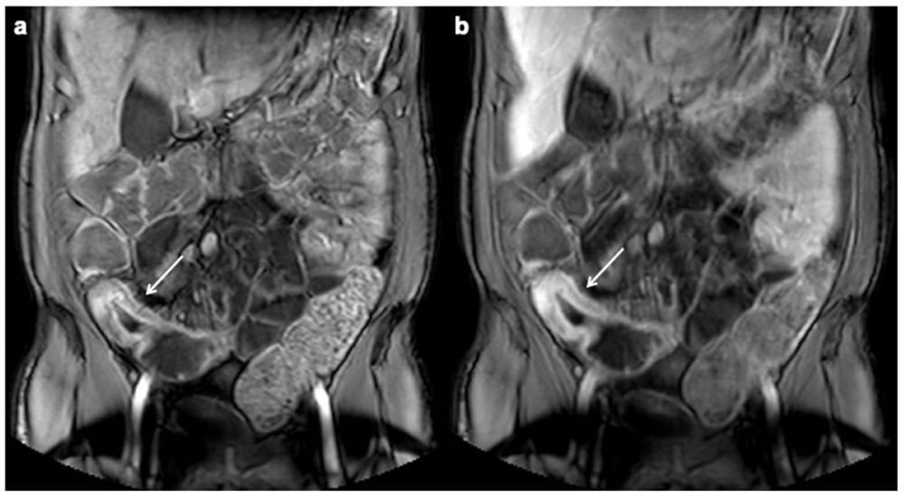
MR Enterography is an imaging exam that will help your physician see detailed images of the small intestine and is commonly used to evaluate conditions such as Crohn's disease, small bowel tumors, and other gastrointestinal disorders. This non-invasive exam visualizes the small intestine's structure and any abnormalities, often by measuring the thickness of the bowel wall.
What to expect on the day of the exam
On the day of your MRI Enterography, you may need to arrive a couple hours early for your oral prep. The MRI scan itself can last from 45 minutes to an hour, but the imaging department may ask you to drink an oral prep an hour or so before imaging.
Why is oral preparation necessary?
The small intestine is 23 feet long and when empty of food and fluid it appears as a deflated balloon when imaged. For better visualization, physicians need to inflate the small bowel and patients may be instructed to drink up to 1500mL (6 cups) of an oral prep between 1 - 2 hours to distend the bowel loops. The volume of oral prep you may be instructed to drink will be determined by your physician.
Your oral prep might be a flavored beverage containing sugar alcohols such as sorbitol and mannitol or a barium sulfate suspension containing sorbitol. The intestines do not easily absorb these sugar alcohols. When ingested, fluids such as water are absorbed by the body, sent to the kidneys, and excreted as urine before it can reach the terminal ileum. This leaves the small bowel poorly distended, not unlike a deflated balloon. The sugar alcohols of the oral prep will help the small bowel to retain fluids and remain fully distended for the imaging exam.
Without adequate distension, the images your physician reads may be less accurate, potentially leading to a misdiagnosis or the need for additional imaging.

Why Bowel Distention Matters
Small bowel imaging requires the intestines to be properly distended to ensure clear and accurate visualization of the bowels. Without sufficient distention, bowel loops can collapse, leading to suboptimal imaging and the potential for missed diagnoses.
Water and milk may seem like easy options for bowel preparation, but they are absorbed too quickly, resulting in poor distention. To counter this, many oral contrast agents use sugar alcohols—such as sorbitol and mannitol—which help draw water into the bowel, keeping it expanded throughout the exam.
The Role of Sugar Alcohols in Bowel Distention
Sugar alcohols, like sorbitol and mannitol, are key ingredients in oral contrast agents because they are osmotically active. This means they pull water into the intestines, helping to maintain the necessary bowel distention for clear imaging. However, there’s a delicate balance—while these ingredients improve distention, excessive amounts can lead to cramping, bloating, and discomfort. In some cases, they may even have a laxative effect, which can be distressing for patients undergoing the procedure.
Some contrast agents contain a significantly higher amount of sugar alcohols than others, which can make the side effects more pronounced. This can lead to increased patient discomfort and, in some cases, incomplete ingestion of the oral contrast, potentially compromising image quality.
A Contrast Agent Designed with Both Image Quality and Patient Comfort in Mind
Your imaging facility may use Breeza® flavored beverage for neutral abdominal imaging as your oral prep. Designed with both the image and patient experience in mind, it provides the optimal level of bowel distention while using a carefully tested balance of sugar alcohols to minimize discomfort.
The radiologist who developed Breeza spent several years perfecting the formulation to achieve the right balance between patient satisfaction and optimal distention. While some degree of cramping or mild laxative effects can occur with most oral prep, Breeza was formulated to reduce these issues compared to competing products with higher sugar alcohol concentrations.
What to expect during the exam
The MRI scan itself typically takes about 45 minutes to an hour. You will lie on a movable table that slides into the MRI machine during the procedure. It is crucial to remain as still as possible during the scan to avoid any motion blur on the images. The machine may make loud thumping or tapping noises, but you will be given earplugs or headphones to help minimize the sound.
After the Exam
Once imaging is complete, you will be able to resume your normal activities. However, be aware that some patients experience limited diarrhea after the exam. If this side effect persists beyond 24 hours or if you feel uncomfortable at any time, contact your physician.

Jonathan McCullough
Product Manager
