
 It’s 7:30 a.m., and the reading list is already building. A breast imaging radiologist logs into the workstation, coffee still warm, and begins scrolling through the first DBT exam of the day. Many more will follow. Each case represents a patient, a story, and the responsibility to detect something that could change, or even save, a life.
But modern breast imaging doesn’t happen in a vacuum. Radiologists today face mounting challenges: rising imaging volumes, physician shortages, and the growing use of advanced technologies like tomosynthesis that improve detection but require more time to interpret. In fact, reading a DBT exam can take roughly twice as long as a traditional 2D mammogram.
At the same time, many radiologists now read for multiple facilities, each with its own workflows, protocols, and documentation styles. The result is a complex diagnostic environment where time is limited and clarity is essential.
Even the Smallest Details can Make a Meaningful Difference
Radiologists rarely meet the patients whose images they interpret. Instead, technologists serve as their eyes and ears: gathering clinical observations, listening to patient concerns, and documenting details that may never appear in the electronic record.
A patient points to a spot of pain.
Another mentions a lump she noticed last week.
A surgical scar sits exactly where subtle distortion appears on the image.
 These details matter.
That’s where something surprisingly simple plays a powerful role: mammography skin markers.
In the white paper, Mammography Skin Markers Transform Uncertainty into Clarity, breast imaging radiologist Dr. Connie Jones, Medical Direct of Breast Imaging at City of Hope Cancer Center, explains how standardized skin marking can dramatically improve communication between technologists and radiologists while enhancing diagnostic clarity.
At first glance, skin markers might seem like a minor detail in an environment defined by high-resolution imaging, advanced software, and artificial intelligence. But when used consistently, they provide radiologists with immediate visual context, helping differentiate benign findings from suspicious ones and allowing them to focus on clinically significant findings.
A Shared Visual Language
Skin markers also create something equally valuable: a shared visual language between the technologist and the radiologist.
A standardized five-shape system helps communicate clinical information quickly and clearly. A pellet identifies the nipple. A circle marks a mole or skin lesion. A line indicates a surgical scar. A triangle signals a palpable lump. A square highlights an area of focal pain or concern.
For a radiologist interpreting hundreds of images in a day, often from multiple facilities, that consistency matters. Instead of guessing what a technologist observed or trying to decipher notes buried in a chart, the information appears directly on the image itself.
Sometimes, that simple marker can prevent a cascade of unnecessary follow-ups.
Research referenced in the white paper found that consistent use of a standardized five-shape skin marker system reduced DBT reading time by an average of 1 minute and 34 seconds per patient, while also improving communication between technologists and radiologists. Over the course of a full reading day, or across thousands of screening exams each year, those minutes add up.
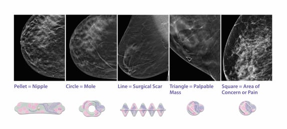
But the real impact goes beyond efficiency. Clear communication reduces ambiguity, decreases unnecessary recalls, and ultimately improves the quality of patient care.
That’s one reason the American College of Radiology recommends using radiographically distinct markers to identify clinical concerns such as palpable lumps, scars, and skin lesions that could affect interpretation.
With increasingly complex imaging technology, it’s easy to assume that innovation always comes from better machines, smarter algorithms, or faster systems. But sometimes the most meaningful improvements are much simpler: a clearly placed marker, a standardized system, and a shared visual language between technologist and radiologist.
Small changes in workflow can reduce uncertainty, improve communication, and make interpretation faster and more confident. And in breast imaging, where every image represents a patient waiting for answers, those small changes can make a very big difference.
Want to learn more about how standardized skin markers improve diagnostic clarity and workflow efficiency? Explore Dr. Connie Jones’s white paper, Mammography Skin Markers Transform Uncertainty into Clarity, to see real-world examples of how consistent skin marking supports more accurate interpretation and better patient outcomes