 The radiologist paused on the image. It wasn’t uncommon. In breast imaging, subtle findings appear every day - areas of density, architectural distortion, or faint irregularities that require careful attention. But this one prompted a familiar question.
"Do we know if this patient had prior surgery?”
As he walked down the hall in the imaging department, the Radiology Supervisor’s ears perked up. It wasn’t a concerning question, but it was a familiar one. Scar tissue can appear on mammography in ways that sometimes mimic suspicious findings. Without clear documentation on the image itself, the radiologist must rely on notes, prior exams, or additional context to determine whether what they’re seeing is expected postoperative change or something that needs further evaluation.
For the imaging leader responsible for the department’s workflow and quality standards, moments like this are important to notice. Not because they signal a failure, but because they highlight one of the subtle challenges of breast imaging: translating patient history into clear imaging context.
Every piece of information that helps the radiologist interpret an image accurately and efficiently, also helps the entire department run more smoothly. And sometimes that clarity depends on something very small.
The Leadership Perspective on Imaging Clarity
Radiology Directors and Imaging Supervisors view breast imaging differently than anyone else on the team. Technologists focus on positioning and patient care. Radiologists focus on  interpretation. But imaging leaders see the entire system: how information moves from the patient to the technologist, from the technologist to the image, and from the image to the radiologist. When that chain of communication works well, everything flows.
But when clinical context isn’t immediately visible on the image itself, small interruptions begin to appear in the workflow. Radiologists pause longer to interpret findings. Technologists may be contacted later for clarification. In some cases, patients may even be asked to return for additional imaging to confirm that what appears on the study is simply a postoperative change.
None of these situations are uncommon in breast imaging. Scar tissue, in particular, is something radiologists evaluate frequently. But from a leadership perspective, each moment of uncertainty represents an opportunity to improve clarity across the department. Because when important details about a patient’s history are clearly communicated on the image, the entire workflow becomes more efficient.
The Challenge of Scar Tissue in Breast Imaging
Post-surgical changes are a normal part of many patients’ breast imaging histories. Lumpectomies, biopsies, and other procedures often leave scars that appear on future mammograms.
To a trained radiologist, scar tissue has recognizable patterns. But when the presence and location of a scar are not clearly identified on the image itself, interpretation can become more complex. A subtle area of distortion may require closer evaluation. The radiologist may compare multiple prior studies. Additional imaging may be recommended simply to confirm that the finding corresponds with a known surgical site.
For the patient, this can introduce unnecessary anxiety. Being called back after a mammogram, even for something benign, can be stressful.
For the imaging department, it adds additional steps to the workflow.
Radiology Directors understand that improving image clarity isn’t only about the technology capturing the image. It’s also about providing the radiologist with the context needed to interpret that image efficiently and confidently.
That’s where a small but powerful solution comes in.
A Simple Marker That Brings Immediate Clarity
When a patient has a known surgical scar, placing a TomoSPOT® scar marker directly over the
site before imaging provides the radiologist with an immediate visual reference point. 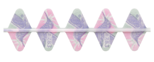
On the mammogram, the marker clearly identifies the location of the surgical scar, allowing the radiologist to correlate the imaging appearance with the patient’s clinical history in seconds.
For the Radiology Director overseeing the department, that small addition has meaningful impact across the workflow.
Radiologists can interpret images more confidently because the location of the scar is clearly documented. Technologists spend less time answering follow-up questions about surgical history. The likelihood of unnecessary callbacks decreases because the radiologist can quickly determine that a finding corresponds with known postoperative change.
And perhaps most importantly, patients benefit from a smoother experience with fewer uncertainties.
Solutions like TomoSPOT scar markers integrate easily into existing workflows. They don’t require new equipment or complicated protocols. Instead, they simply ensure that important clinical context travels directly with the image.
For imaging leaders focused on quality and efficiency, those kinds of improvements are invaluable.
When Small Improvements Strengthen the Entire Department
Later, when the Imaging Manager reviews departmental performance, the success of the breast imaging program is measured in more than just the number of exams completed. It’s reflected  in the clarity of the studies being interpreted, the efficiency of the radiologists’ reading workflow, and the confidence patients feel in their care.
These outcomes rarely come from a single major innovation.
Instead, they come from dozens of thoughtful improvements that remove small barriers from the imaging process, tools that help technologists capture better clinical information, processes that support radiologists during interpretation, and solutions that ensure nothing important gets lost between the patient and the image.
TomoSPOT skin markers represent one of those simple but meaningful improvements. By clearly identifying nipples, scars, skin lesions and areas of concern on mammography images, they help radiology teams interpret studies more efficiently while reducing unnecessary uncertainty.
For imaging leaders responsible for delivering high-quality breast imaging every day, that kind of clarity makes a difference and sometimes the smallest tools make the biggest impact. To hear more about imaging workflow improvements from a Radiologist’s perspective, check out our complimentary CEU, Architectural Distortion from Surgical Scarring in 3D Mammography.