
 At 7:10 a.m., the mammography supervisor unlocks the imaging suite. The first patient isn’t scheduled until 7:30, but the day has already started in her mind. She scans the schedule: a full morning of screening exams, a few diagnostic add-ons squeezed into the afternoon, and a biopsy scheduled before the end of the day. One technologist is still getting comfortable with positioning, and another called out sick.
It’s a familiar rhythm in breast imaging, one that requires constant attention to both the big picture and the smallest details. Because behind every exam, every image, and every patient interaction sits a single, critical question: Did we capture everything we needed to see?
A Knock at the Supervisor’s Door
The first patient of the morning arrives for a diagnostic mammogram. Not long after, a technologist lightly knocks on the supervisor’s office door. “Can you take a look at something?” she asks.
Inside the exam room, the patient stands at the mammography unit, shoulders tense, eyes focused on the floor. A prior mammogram showed an area of concern, and today’s images will help determine the next step. The technologist gestures toward the screen and explains that there’s a mole very close to the area they’re evaluating.
It’s a situation mammography supervisors recognize instantly. Small details on the skin can easily create big questions on an image. Is the density real? Is it something on the skin surface? Will the radiologist question it and request additional views?
Every moment of uncertainty has consequences. The technologist may need to take extra images. The patient may wait longer for answers. Anxiety can quietly build in the room.
Much of a mammography supervisor’s role is about removing exactly that kind of uncertainty.
Throughout the day, they guide technologists through positioning challenges, troubleshoot image questions, and ensure the team has the right tools to get it right the first time. In this moment, the solution is simple.
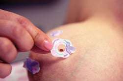 The technologist reaches for a small adhesive marker and gently places it over the mole before capturing the next image. The marker, a TomoSPOT, will communicate the skin lesion by its mere presence on the image. When the image appears on the screen, the difference is immediate.
What could have been an ambiguous density is now clearly marked as a mole. The radiologist will immediately understand what they are seeing. No guessing. Just clarity.
Smoother, More Confident Imaging
Skin markers may seem like a small part of the imaging process, but mammography supervisors know how much influence they can have on the entire workflow. Without clear skin marking, radiologists may question whether a density originates from the skin or from deeper breast tissue.
That uncertainty can quickly lead to additional views, longer exam times, diagnostic delays, and increased anxiety for the patient waiting for answers. Supervisors understand the ripple effect.
One small question on an image can add minutes to an exam, create delays for the next patient, and leave technologists feeling rushed. But when skin findings are clearly marked, the entire imaging process becomes smoother and more confident for everyone involved.
By mid-morning, the department is moving quickly. Patients move through the waiting room.png?width=296&height=207&name=BeekSpeak%20Blog%20Body%20Image%20(29).png) and into exam suites, technologists rotate between rooms, and radiologists review images in real time. The supervisor checks in with the team as she walks the hallway, making sure everyone has what they need to keep the day running smoothly.
and into exam suites, technologists rotate between rooms, and radiologists review images in real time. The supervisor checks in with the team as she walks the hallway, making sure everyone has what they need to keep the day running smoothly.
She knows her technologists are skilled and deeply committed to their patients, but even the most experienced professionals benefit from tools that make their work clearer and easier.
That’s where TomoSPOT skin markers quietly support the workflow. They are simple, quick to apply, and highly effective at communicating critical information directly on the image. In a field where clarity matters so much, that small detail can make a meaningful difference.
Simple Tools to Boost Communication
As the afternoon unfolds, the team completes dozens of exams: some routine, some diagnostic, and a few that require extra care and attention. The day has been busy, but the workflow has stayed steady.
There are fewer questions from the interpreting physicians, fewer extra images needed to confirm findings, and fewer moments where technologists pause to second-guess what they’re seeing. And it didn’t happen because of a major technological breakthrough or a dramatic process overhaul. It happened because the team used a simple tool to boost their communication – skin markers.
Mammography supervisors understand this better than anyone. Their responsibility extends beyond schedules and staffing. They support technologists, protect the patient experience, and help ensure that every image tells the clearest possible story.
That’s why even small improvements matter. A clearer image. A faster exam. A more confident interpretation. Sometimes all it takes is a small marker placed in exactly the right spot. In mammography, small changes truly can make a big impact.
If you’re a mammography supervisor focused on improving workflow, image clarity, and patient experience, we invite you to view our on-demand webinar to explore how small improvements - like the effective use of skin markers - can make a measurable difference in your daily imaging workflow.