
Skin marking in MRI has evolved over the years in an attempt to keep up with the technology it supports. With recent advancements in hardware and improved sensitivity of surface coils, imaging in a small field of view is producing images with exquisite anatomical detail.
Challenges when limiting the field of view to a smaller area
Similar to the challenge technologists experience when trying to communicate a level on T-Spine studies, it is sometimes difficult to orient certain studies when telltale anatomy is eliminated from the image.
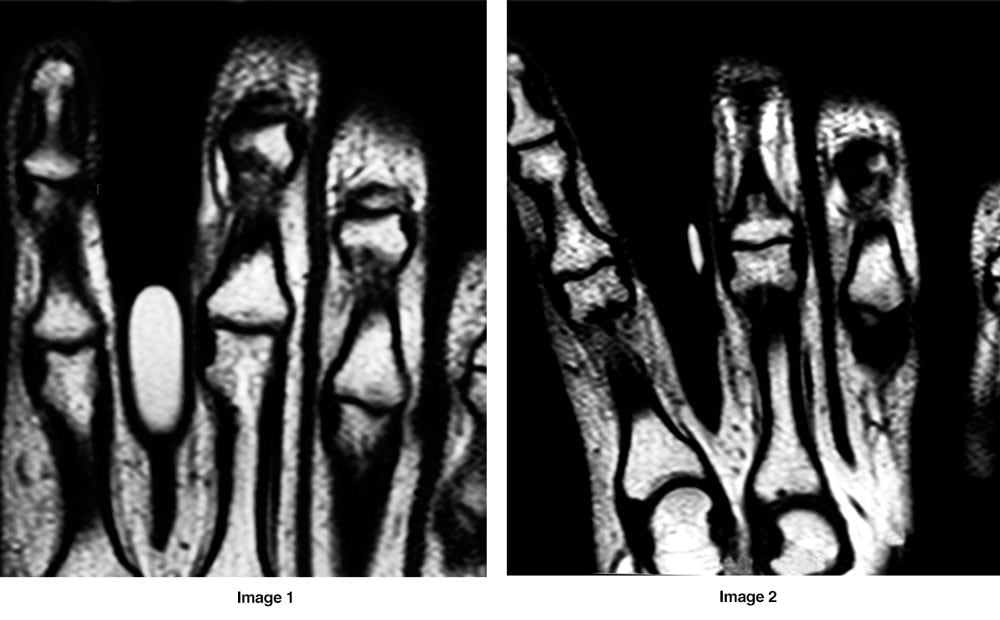
Fingers can fall into this category, especially when imaging the PIP joints or the thumb as seen in the images below.
In Image 1, it is difficult to tell which finger the cod liver pill is marking, or exactly where along the finger the point of pain is located.
However, in Image 2, a smaller, more specific marker (OrthoSPOT Packets™) is used and it is clear which is the affected finger and the PIP joint is localized as the area of concern.
Specificity matters
When trying to communicate the location of a palpable mass or a point of pain it is very helpful to be as specific as possible.
Wadhwa, et al. looked at the correlation between skin marker placement and the detection of pathology. They concluded that, “Skin marker placement can aid in detection of clinically important imaging findings”
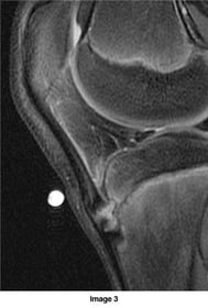 The marker in Image 3 was placed at the point of focal pain and is directly adjacent to thickening of the distal patellar tendon consistent with early Osgood Schlatter disease.
In addition to being helpful when pathology is present, radiologists also find smaller markers helpful when used on an exam that is read as negative.
The presence of the marker gives the radiologist confidence that they are evaluating the correct area, especially when the patient history is vague.
Marker size matters
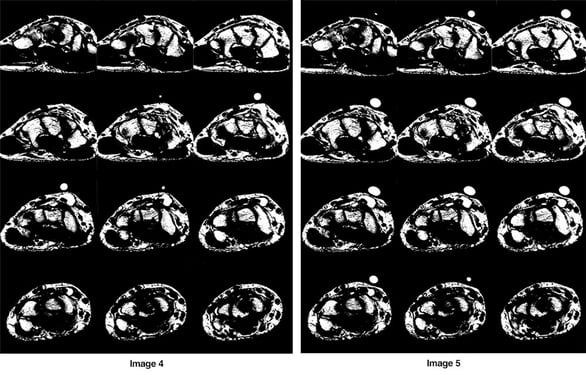
The marker in Image 4 (PinPoint® for small field of view imaging), has a 6mm internal diameter, and depending on slice thickness, will image on two to four slices.
In this example, a ganglion cyst is obvious. But when pathology is subtle, or the exam is negative, the presence of the marker on fewer slices increases accuracy.
In comparison, the cod liver capsule in Image 5 appears on 11 of 12 slices.
Communicating small surface findings that could indicate hidden pathology
 In this case of suspected spina bifida (Image 6), the marker is used to identify the location of a surface finding which will help differentiate between an innocent coccygeal dimple and a dermal sinus tract (DST).
It is also common practice to place a marker at the site of ulceration in the case of suspected osteomyelitis. Shoots, et al., suggest the following when evaluating the diabetic foot with an ulcer:
“To determine whether osteomyelitis is present, place a marker on the ulcer or sinus tract and track it down to the bone and evaluate the MR- signal intensity of the marrow”.
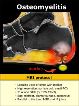
For all of these applications it is important to use a marker that will communicate the specific area of concern as this may aid in dictation and reduce reading time.
Skin markers designed for small field of view imaging in MRI
PinPoint® for small field of view imaging is perfect for communicating a specific point of pain, palpable lump, or other area of concern. The internal diameter of 6mm ensures the marker will only image on 2-4 slices.
At 7.5mm, OrthoSPOT Packets™ is also useful in these situations and it has the added benefit of a low profile that will eliminate indentation when used inside a tight dedicated extremity coil.
Visit beekley.com or call your 1-800-233-5539 to learn more about Beekley's line of professional skin markers for MRI.
For product safety information, visit Beekley.com
Related articles:

Richard Foster
Director of Training
