While every team member in radiation oncology is vital to patient care, it is the role of the medical dosimetrist to prepare the treatment plan and make sure the plan will work as designed: deliver the most effective dose of radiation to kill the cancer with the fewest side effects to the patient’s healthy organs and tissue.
As you can imagine, accuracy in calculating the plan is critical.
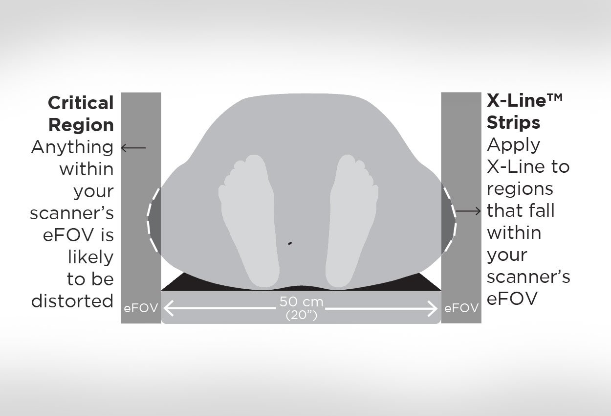
One factor that can negatively affect accuracy is when any part of the patient's body contour falls outside the scanable field of view.
America's Growing Girth Problem
Increasingly, Americans are outgrowing technology and not in a good way. According to a 2017 data brief put out by the Center for Disease Control/National; Center for Health Statistics, 39.8% of adults (roughly 4 out of every 10) are considered obese. Lifestyle choices and socio-economic factors aside, obesity in and of itself is a risk factor for several diseases, including cancer. As a result, the imaging and radiotherapy communities are dealing with the challenges of providing care to this growing population.
The good news is equipment manufacturers have responded with imaging equipment that can accommodate larger patients. For example, Toshiba's Aquillion™ LB was designed with bariatric patients in mind and boasts a 90 cm gantry opening with a 70 cm true field of view and an 85 cm extended field of view (eFOV).
The bad news is that big equipment costs big money and may require big changes within the facility to even have on premise. So there are many clinics that may not have access to more modern equipment designed to meet the unique needs of treatment planning for bariatric patients and as a result, there are times when patient anatomy extends to and beyond the extended field‐of‐view.
 "Confounding Artifact"
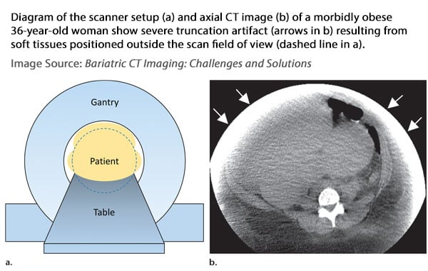
In their paper "Bariatric CT Imaging: Challenges and Solutions," Fursevich et al. state that "images of bariatric patients are susceptible to confounding artifacts." Image noise, truncation, and cropping are among the most common cited in their paper.
One method that is commonly employed to deal with the issue of image truncation involves image fusion of overlapping regions of 2 or 3 scans with the center of each scan set at different positions within the patient. The drawback to this method is that it requires quite a bit a physical exertion to reposition the patient between scans, is difficult to replicate positioning with each shift, and, of course, exposes the patient to additional radiation exposure.
Alternate methods involve either manual or auto removal of artifact to help calculate the body contour that is missing from the image. However, whether it's done by a professional or AI, it's still just a best estimate of where the skin line actually is in the extended field of view.
"Dosimetrists the Champions"
When it comes to IMRT, knowing where the skin line is is crucial according to Tina Voytek, the Chief Radiation Therapist for cancer center in Northern Virginia.
IMRT (Intensity-modulated radiation therapy) is an advanced mode of radiotherapy requiring high precision in treatment planning. It allows for the radiation dose to conform more precisely to the 3-D shape of the tumor by modulating the intensity and shape of the radiation beam in multiple small volumes. This precision also minimizes the dose of radiation delivered to surrounding normal critical structures.
 Tina's facility does a lot of IMRT and she was intrigued when she saw X-Line™ body contour tape for radiotherapy on the Beekley Medical Website. When applied to regions of the body that fall into the eFOV, X-Line's three radiopaque lines appear as dots, making it easy to identify the skin line and true body contour.
Although it's not often that her patients fall into the eFOV (approximately 1 or 2 a month), Tina told us that when it did occur, dosimetry would either have to rescan the patient or estimate the external body contour. She shared the user guide with her physicists and dosimetrists and they were excited to try X-Line to see how it could save them time and improve accuracy in those situations.
Tina told us that the dosimetrists were the champions of the purchasing decision of X-Line after their evaluation as they found it to be more accurate and helped with their planning as they could now visualize the whole contour of the body.
Since purchasing X-Line, Tina's team uses it proactively on any tissue that falls into the extended field of view - shoulder to wrist, upper arms, prone or supine pelvis, abdomen, really anywhere the body overlaps the table.
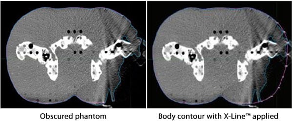
"Just Show Them the Phantom!"
When I asked Tina what advice she would give to her peers interested in evaluating X-Line, she told me "Just show them the phantom!"
Who knows - if you take her advice, you might just become your dosimetrist's new best friend.
To learn more about X-Line, contact your Radiation Oncology Business Development Manager at 1-800-233-5539 or email info@beekley.com
Related articles:

Mary Lang Pelton
Director of Marketing Communications
