A mammographer’s goal is to produce the best clinical images while providing the patient with a positive experience. The perfect image combines the science of imaging technology and the art of positioning.
Mammography is considered one of the most challenging modalities within medical imaging. Technologists have many factors to think through when positioning the patient – body habitus, breast composition and pliability, physical limitations or ailments, as well as the patient’s emotional state.
The Facts – Positioning is the #1 Failure during ACR accreditation
In 2015, the American College of Radiology (ACR) found that of all the clinical images which were deficient on the first attempt at accreditation, 92% were deficient in positioning. In facilities that were already ACR-accredited, 79% of all unit accreditation failures were also due to positioning.
Mammographers report that between 15% and 40% of patients are difficult to position and even though they may have limitations that would make the perfect image almost impossible to achieve, the goal is to get as close to perfect as possible.
When considering the MLO view the following criteria apply:
- Pectoralis muscle is well visualized
- Muscle is wide superiorly
- Down to the level of the PNL
- Convex or straight anterior border
- Retroglandular fat is well visualized
- Nipple is in profile
- IMF is included and open
When speaking with mammography supervisors across the country about the FDA’s EQUIP initiative, the one area that many of their technologists find challenging is the IMF positioning requirement. The IMF needs to be included on the image, it has to be open, and without vertical or horizontal skinfolds.
The Fix
When discussing a fix for this problem many technologists attribute the lack of an open IMF to an inadequate “up and out” maneuver. And while this may be part of the problem it is usually not the entire problem.
According to Shelly Lille’, B.S., R.T.(R)(M) and Wendy Marshall R.T.(R)(M)(QM), both positioning experts and the authors of "Mammographic Imaging: A Practical Guide", the path to an open IMF is intertwined with every other MLO positioning consideration.
They suggest the following steps to ensure that the IMF is acceptable:
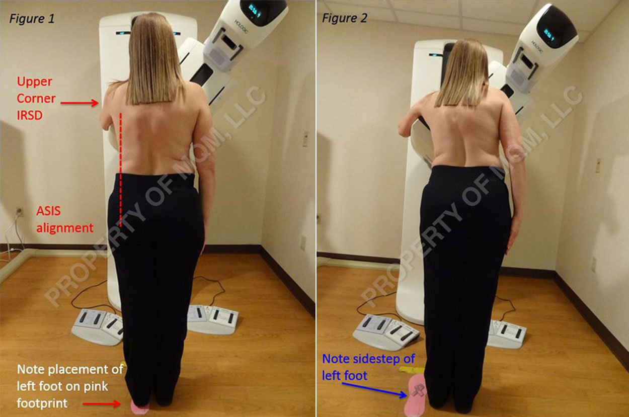
Begin with the patient’s feet and pelvis directly facing the gantry. Then sidestep the patient in order to align the ASIS with the inferior corner of the IRSD; this action engages the IMF region to the front of the IRSD. (Figure 1 - INCORRECT feet placement, Figure 2 - CORRECT feet placement).
Ensure the upper corner of the IRSD is placed just anterior to the Latissimus Dorsi muscle and that the entire thorax from this point downward to below the IMF region is in intimate contact with the chestwall edge of the image receptor. For sufficient inclusion of the retroglandular fat space, the chestwall edge of the IRSD should contact the thorax approximately 1.5-2 inches behind and parallel to the mid-axillary line.
Leaning the patient onto the IRSD for the MLO projection in order to place the axilla at the superior chestwall corner (specifically just anterior to the Latisimus Dorsi muscle), naturally introduces a “buckle” in the lower region of the thorax.
This “buckle”, introduced near the IMF, can be eliminated by bending the ipsilateral knee and ensuring that the pelvis is slightly shifted away from the IRSD (the lower thorax should remain in contact with the IRSD).figure 3 This positions the sternum approximately parallel to the IRSD.
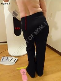 Bend the ipsilateral knee and slightly shift the pelvis away from the IRSD - the lower thorax should remain in contact with the IRSD as seen in Figure 3.
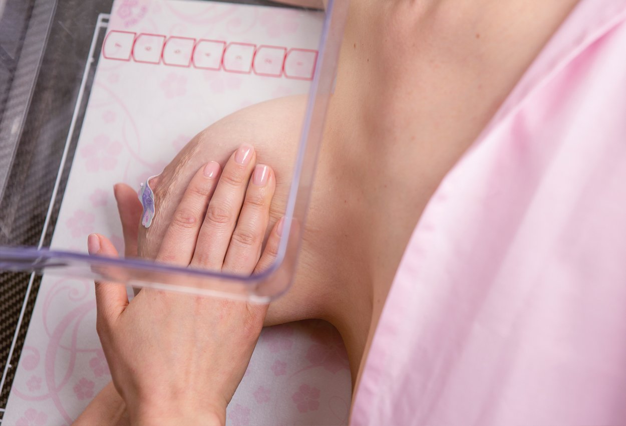
With a flat hand, begin at the axilla and sweep the lateral breast skin in a downward direction, following this action through and past the IMF region. This tissue sweep of the IMF region ensures intimate contact between lateral tissue and IRSD and between medial tissue and compression device. Tissue sweep also ensures elimination or reduction of skinfolds.
Ensure posterior breast tissue has been secured on the IRSD and continue to coach the patient to “completely relax”, or to “melt or collapse” onto the IRSD.
Support the breast tissue outward approximately 90o from the thorax with assurance that medial breast tissue is not allowed to droop.
Apply adequate compression to keep the breast in place and maintain an open IMF.
The Mammographer's Helping Hand
In addition to these steps there is clinical evidence that the use of Bella Blankets® protective coverlets for mammography can assist with the acquisition of an open IMF.
In a 2013 study performed at a dedicated breast center, images from the prior year where no coverlets were used were compared to the current year where Bella Blankets were used. The IMF as seen as “open” improved from 67% to 87% with the addition of Bella Blankets.
This improvement was attributed to the ease with which the mammographer can keep the breast 90o to the thorax without the breast drooping resulting in a closed IMF.
“Bella Blankets’ texture keeps breast tissue from sliding back and it’s easy for the tech to get her hand out from under the breast without losing breast tissue. We documented more tissue YOY in both CC and MLO views during our trial period. We liked the ability to get the maximum tissue on each image; no skin folds or wrinkles, and the IMF is open.”
~Manager Breast Center & Imaging Center, Memphis, TN
Learn what other customers are saying about how Bella Blankets help both clinically and with patient satisfaction. Download the Bella Blankets® Customer Testimonials booklet or contact Beekley Medical at 1-800-233-5539 or info@beekley.com about a trial evaluation to experience how Bella Blankets can help you with the elusive inframammary fold.

