
Are you experiencing a blurring or slinky artifact with your skin markers in digital breast tomosynthesis (DBT)?
Using skin markers intended for 2-D mammography could be your culprit.
DBT or 3-D mammography is very sensitive to densities – this is what makes it so great at detecting cancers, however, not so great when the dense object is a skin marker designed for optimal imaging with older technologies.
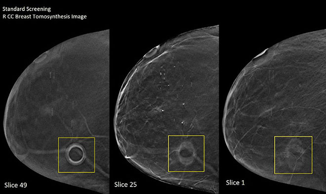
Figure 1
Breast tomosynthesis image – Patient A – standard screening right CC view. A commercially available mole marker with a higher density inner ring surrounded by a lighter density outer ring. (A) Slice 49 shows the marker at its clearest resolution. (B) Slice 25 shows the marker beginning to fade and artifact. (C) Slice 1 the marker artifacts and continues to image on the last slice.
A skilled radiologist who appreciates the value of skin markers in communicating breast topography (moles or scars) and patient complaints (palpable lumps, points of pain) can learn to “work around” the artifact, but read time suffers and there is always a chance of requesting additional views if the artifact is obscuring tissue detail.
A useful tool shouldn’t be something you have to “work around.” That kind of defeats its usefulness.
However, what if there were a mammography skin marker specifically designed, tested, and proven to image with minimal or no artifact in 3D mammography?
A skin marker calibrated for the 3D scanning portion of DBT can help reduce your read time and those additional views resulting from obscuring skin marker artifact.
A thinner marker made with a uniform low-density created for optimal imaging under the 3D scanning portion of DBT will result in significantly reduced artifact over a thicker marker with differing densities originally intended for 2D imaging. .
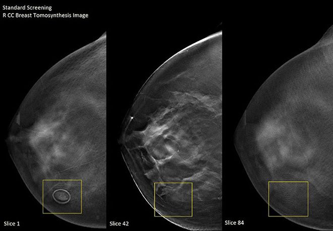
Figure 2
Breast tomosynthesis image – Patient B – standard screening right CC view. (A) TomoSPOT® (Beekley Medical) mole marker consisting of a single made from a single, uniform low-density ring appears in slice 1 in clearest resolution. (B) Slice 42 reveals that the marker has faded with the mole. (C) Slice 84 shows minimal “slinky” artifact.
In a double-blind side-by-side comparison, 32 radiologists were asked to examine the images shown in Figure 1 and Figure 2 and indicate which mole marker they preferred and why.
More than 93% of the radiologists preferred mole marker B over mole marker A.
- “[Marker B] does not obscure underlying skin lesion and does not appear as a breast density on other images.”
- “[There is] less artifact obscuring adjacent tomo slices.”
- “No slinky effect.”
- “Less distortion of the breast tissue on subsequent images.”
- “[The] image appears less distorted/blurry in marker B image than in A.”
As technology evolves, so must your everyday tools. If you’re tired of “working around” your skin markers’ artifact in Digital Breast Tomosynthesis, request a complimentary trial of TomoSPOT skin markers for digital breast tomosynthesis and see the difference for yourself.

