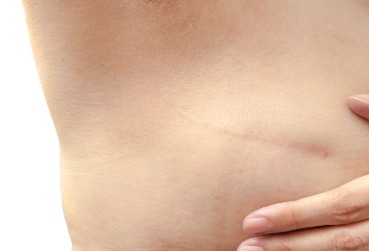
Dr. Douglas Housman is a radiation oncologist previously with Memorial Sloan-Kettering in NY, and now at the Harold Leever Regional Cancer Center in CT. In his many years of experience, there’s one thing he can attest to: you can’t always see scars in CT images during treatment planning.
Dr. Housman explains how he overcomes this challenge.
“When we’re planning the treatment path, it’s optimal to know where the skin projection and scar are in relation to the lumpectomy bed. We use (topical radiopaque) markers to indicate scars and delineate superficial landmarks like the areola.”
But why is it hard to see the scar in the CT image? Part of this is due to one of the most positive advances in treating breast cancer – oncoplastic surgery.
Greater Accuracy and an Oncoplastic Approach
Oncoplastic breast conservation is a surgical approach that not only removes the cancer but is focused on maintaining the natural look and feel of the breast and prevent excessive scarring.
Dr. Housman explains how this has changed treatment planning.
“More physicians are taking an oncoplastic approach to treating breast cancer,” he says. “Gone are the days where an oncologist would treat the tissue within a 2cm margin of the scar bed. What our team does now is evaluate the 3D scan and contour a clinical target volume. It’s more accurate, but it’s not just about accuracy.”
While oncoplastic surgery is a great value to the patient, it can cause internal scarring. This is what makes it more difficult to read CT images and accurately identify the lumpectomy bed.
“When you take an oncoplastic approach, it utilizes techniques from plastic surgery. This often manipulates and reapproximates the tissues past the surgical recession. Therefore, the tissue defect is lessened after a lumpectomy, but your target is very different.”
You Can’t Hit a Target You Can’t See
 Because of the internal scarring and change in tissue, it can be hard to hit a target that you can’t see.
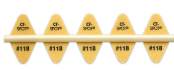
“This is where the CT-SPOT® line is helpful. It’s hard to delineate scars and superficial landmarks when you can’t see the scar in CT. A low artifact radiopaque wire to identify the lumpectomy bed or areola just makes treatment planning so much easier. Our dosimetrists really appreciate it.”
“It’s especially useful when we do our boost treatment planning. We utilize the 3D data obtained from the CT simulation and verify the superficial landmarks and scars on the skin projection.”
To learn more about CT-SPOT® line, contact your Radiation Oncology Business Development Manager at 1-800-233-5539 or email info@beekley.com. Visit Beekley.com for product safety information.

