Mammography technologists are the eyes and ears of the radiologist; which makes them critically important in the breast imaging process.
Aside from the performance of high quality imaging, the most important service that a technologist can provide for their patient is effective communication.
From the moment a woman walks into a breast imaging facility until the time she leaves she will likely have angst about whatever examination that she is there to have performed.
Whether it is for a screening mammogram or a symptomatic diagnostic examination, everything about a woman’s experience in the radiology department is heightened because of the fear of breast cancer: “Will this be the day that I am told that I have breast cancer?”
Effective, concise and accurate communication (and education) of the patient can significantly lessen her anxiety and improve her experience.
It can also improve the quality of her examination because she is more relaxed and able to more fully cooperate with the examination or procedure.
To that end, the communication skills of the technologist play an important role in patient experience since the technologist will see all of the patients (the radiologist usually only sees the diagnostic patients).
Part of effective communication involves thorough and accurate history taking.
The technologist literally serves as the gatekeeper, making sure that the correct examination is performed based on the patient’s presentation.
A screening patient should be asymptomatic or have symptoms that are longstanding or have been worked up previously and found to be benign.
Diagnostic patients may be diagnostic based on symptoms that the patient perceives, physical findings that the referring clinician perceives, or based on a past history that warrants a problem solving evaluation (such as lumpectomy for malignancy).
Accurate and detailed history taking in this instance ensures that the correct examination(s) are performed for that particular patient making the likelihood of a correct diagnosis more likely.
The technologist’s familiarity with the specifics of these criteria is crucial to the accuracy of the information.
Speaking the same language avoids pitfalls in understanding
 In The Breast Test Book: A Woman’s Guide to Mammography and Beyond, Doctor Michael Linver and I provide an easily understandable narrative of the distinct differences between screening and diagnostic patients.
The chapter on “Screening the Asymptomatic Population and Saving Lives” and the chapter on “Symptomatic Patients and the Journey to the Correct Diagnosis” would both be useful review for any technologist who performs breast-imaging examinations.
Now that the technologist has acquired the appropriate history from the patient, the next step is to accurately document and communicate the relevant information to the radiologist.
This step is necessary to ensure that all data that would affect the outcome of the examination (the radiologist’s assessment as to the benignancy or suspicious nature of the evaluation) is communicated to the radiologist.
In this instance, speaking the “same language” is important so that the information conveyed is interpreted with the same level of understanding between the two parties that are communicating.
As stated in the Glossary 1 (Terminology 101) in The Breast Test Book: A Woman’s Guide to Mammography and Beyond, the terms used within breast imaging help to succinctly and effectively communicate information.
Based on agreed upon conventions, certain words and phrases have a specific meaning within the context of a particular field. There is no need for exhaustive explanation because everyone knows what that term means and what it implies when used by practitioners.
Translating what patients say into what they mean
An example of this would be the terms excisional biopsy versus lumpectomy.
Excisional biopsy is defined as “surgical removal of a portion of the breast that includes the abnormality or region of interest; typically refers to removal of a benign lesion.” Lumpectomy, on the other hand, is defined as a “surgical procedure to remove cancer or other abnormal tissue; implies that the final pathology result was cancer.”
It is the responsibility on the technologist to clarify with the patient what she means by “lumpectomy” - which is a common description by patients who have had both benign and malignant surgical procedures.
Whether the patient has had a previously documented cancer will affect the radiologist’s interpretation and may explain other findings such as thickened skin or trabecular thickening that can be seen in lumpectomy patient’s who have also had radiation therapy.
Visual communication with skin markers assists with interpretation and clinical efficacy
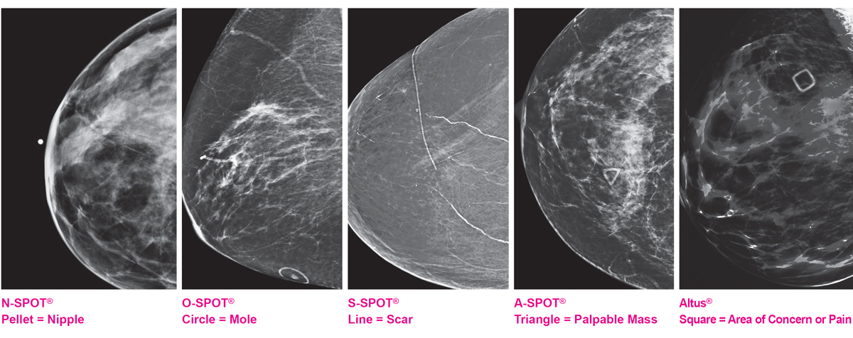
Another effective way for technologists to communicate with the radiologist is via the use of skin markers.
Skin markers aid the radiologist by providing a visual form of communication. By following a consistent protocol for the use of skin markers, it can effectively communicate: nipples in profile, moles or raised skin lesions, scars, palpable masses and areas of concern.
If specific protocols for skin markers are consistently used within a practice, the image interpretation for the radiologist becomes more accurate and efficient; which is of course better patient care.
Beekley Medical developed the 5-Shape Skin Marking System for Mammography which consists of: A pellet marker for indicating nipple location, a circular marker for indicating a raised mole, a linear marker for indicating post-surgical scars, a triangular marker for indicating a palpable finding, and a square marker for indicating non-palpable areas of concern or pain. These specific shapes help to effectively communicate to the radiologist at a glance.

According to mammography supervisors, the top reasons for using skin markers are:
- To clearly identify skin lesions and skin surface abnormalities
- To provide an accurate method of communication to the radiologist
- To aid the radiologist in diagnosis
- To highlight nipples, moles, scars and masses
- To avoid unnecessary callbacks and to reduce recall rates
Channeling what you do every day to improve communication results in better patient care.
Mammography technologists are uniquely positioned to act as the conduit of communication and transference of knowledge between patient and radiologist.
Patients are often anxious at their exam due to the prospect of "something" being found on their mammogram. This anxiety is often heightened by a lack of knowledge about what to expect from the process of breast imaging evaluation.
Physicians often encounter patients who have little or no understanding of the reasoning behind the examination or procedure about to be performed - sometimes even up to the day of their breast cancer surgery.
The fact remains that the majority of women undergoing breast evaluations will not be diagnosed with cancer. However, symptoms of benign breast abnormalities are quite common and impact many more lives. Accurately diagnosing and explaining these non-cancerous conditions can alleviate much anxiety, in addition to helping patients towards a correct treatment plan.
I invite you to review, The Breast Test Book: A Woman’s Guide to Mammography and Beyond (Oxford University Press, 2017) for more helpful tips and information that can help you serve as a better and more effective communicator for your patients and your radiologists.
Related articles:

Connie Jones, M.D., Fellowship Trained, Breast Imaging Radiologist, Author of: “The Breast Test Book: A woman’s guide to mammography and beyond”
