
Artifact can be an unavoidable obstacle for the radiation oncology team. In radiation therapy, no one knows this more than the medical dosimetrist who is responsible for interpreting the images taken during CT simulation and determining the appropriate dose and dose distribution.
In many cases, artifact images as light streaks across normal tissue. This can distort the true density of the normal tissue (measured by houndsfield units), and put the patient at risk of receiving either too little or too much of a radiation dose.
Advances in technology for removing artifact in treatment planning
Software has been developed to reduce artifact and noise, while other medical professionals have developed techniques to work around it, such as image reconstruction and utilization of images from other modalities.
Still, dosimetrists resort to auto-contouring images - matching the affected tissue with the houndsfield units of nearby tissue unaffected by the streak artifact.
This tedious and time consuming task must be performed on every slice on which the artifact appears for that patient. A miscalculation in the dose could harm nearby healthy tissue or prove to be less effective on the cancer cells and tumor growth.
What causes artifact?
Most streak artifact occurs near high attenuation materials, such as bone or metal, and tends to be the result of beam hardening and scatter.
In many cases one can eliminate some of this artifact by evaluating the tools used in treatment planning. High density skin markers are prone to creating such streak artifact.
However, skin markers remain an important tool and valuable guide for those involved in treatment planning, particularly for 3-point set-ups, isocenters, field borders, and other specific points of interest.
Low density skin markers can help reduce streaking
Beekley Medical®’s CT-SPOT® skin markers are designed to reduce and minimize artifact in CT treatment planning.
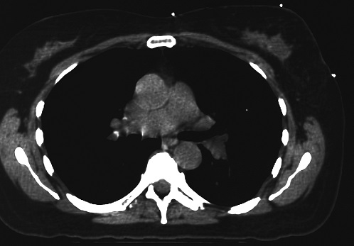
Axial slice showing 3-point setup of breast using CT-SPOT® pellet markers.
Note the crisp. clear imaging of the 3 markers used and absence of streaking artifact emanating from the areas marked.
These non-metallic pellets, line, and crosshair skin markers image brightly on every slice and help to reduce the time the dosimetrist spends autocontouring while remaining a visual aid for the interpreting radiation oncologist.
Learn more about how Beekley Medical can help you achieve greater precision, accuracy, and patient care:
Contact your Business Development Manager at 1-800-233-5539 or +1-860-583-4700 outside the United States to request your copy of Solutions to Daily Challenges in Radiation Oncology or click here to download it now.

