Can the type of skin marker used make a difference in revealing potential breast cancers? While there is no substitute for the trained eye of a radiologist who specializes in breast imaging, the type of mammography skin marker used to identify a post-surgical scar, raised mole, palpable mass, or non-palpable area of pain or concern can either help or hinder when it comes to spotting microcalcifications - important, early indicators of new or recurrent breast cancers.
Because the number, size, shape, and distribution of microcalcifications seen in breast tissue can make the difference between a benign or potentially malignant interpretation of the breast image, it is crucial that they be viewable through any skin marker.
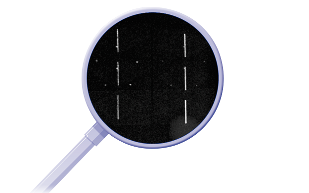
You might think it's the size or width of the marker that can obscure tissue detail, but in all actuality, it's the density of the marker that makes the difference when it comes to revealing potential breast cancers.
The smallest detectable microcalfication in mammographic imaging is .2–.3mm. Despite being extremely thin, a high-density, 1mm metallic skin marker has the potential to hide this important information from the radiologist's eye.
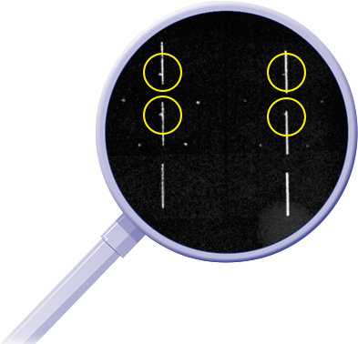
Image 1 – 1mm diameter high-density scar marker on phantom image over calcifications
Although thicker in diameter, mammography skin markers made with a uniform low-density nonmetallic material allow for maximum visibility of underlying tissue detail right through the marker,making it easier for the radiologist to detect breast cancer in it's earliest, most treatable form.
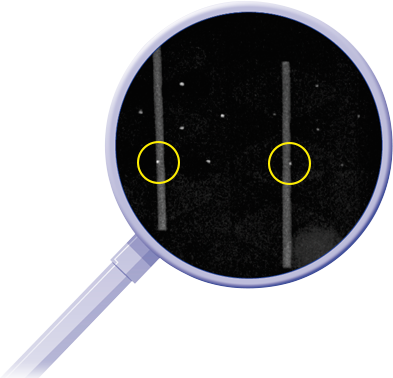
Image 2 – 3mm diameter low-density scar marker on phantom over calcifications
 Click image to read why radiologists feel opaque skin markers most likely to obscure calcifications.
Related articles:

Mary Lang Pelton
Director of Marketing Communications
